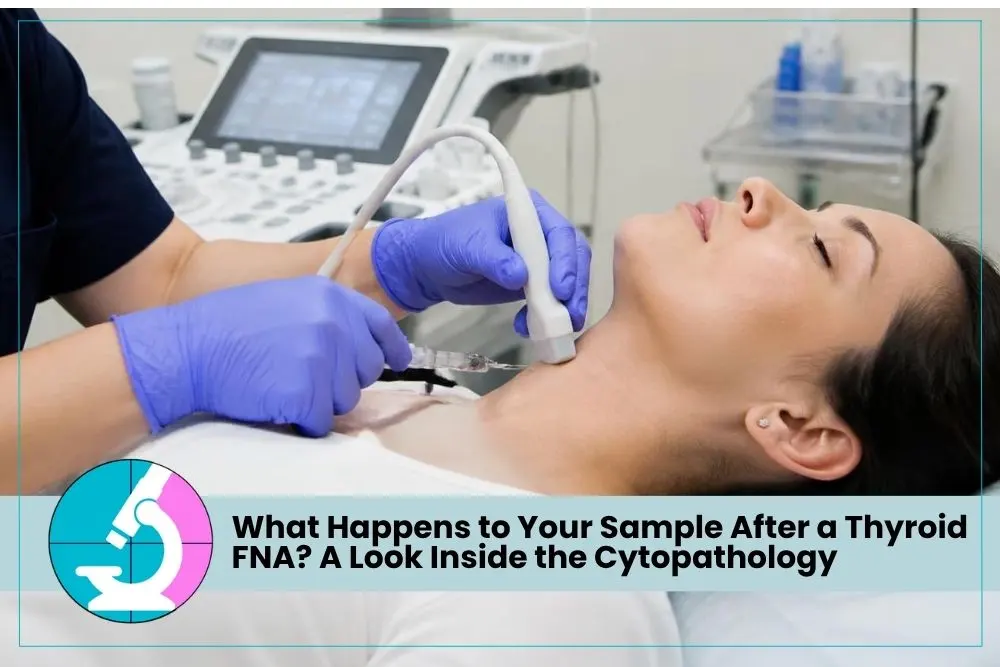

What Happens After a Thyroid Fine Needle Aspiration Biopsy?
When patients come to my office for a thyroid Fine Needle Aspiration biopsy, most of their questions understandably focus on the procedure itself. Will it hurt? How long will it take? Can I go back to work afterward?
But there is another part of the process that patients rarely get to see: what happens to those tiny samples after they are collected.
Once a thyroid FNA sample is collected, it begins a careful journey through the lab long before a diagnosis is reached. My FNA biopsy page explains what the procedure itself involves.
The sample may be small, but it contains an enormous amount of information. The cells collected from a thyroid nodule can help us determine whether the nodule is benign, suspicious, malignant, or requires further evaluation.
As a cytopathologist and Fine Needle Aspiration specialist, I have a unique perspective on this process because I perform the biopsy and evaluate the cells myself. There is no separation between the physician collecting the sample and the physician looking at it under the microscope.
For patients, understanding what happens behind the scenes can make the thyroid biopsy process feel less mysterious. There is much more happening between the moment the needle leaves the nodule and the moment you receive your results.
How a Thyroid FNA Sample Is Collected and Prepared
The quality of a diagnosis begins with the quality of the sample.
Before performing a thyroid nodule biopsy, I use ultrasound to examine the nodule and determine exactly where I want to collect cells. Thyroid nodules are not always uniform. Different portions can have distinct characteristics, making precise targeting important.
A very thin needle is then guided into the nodule to collect cellular material.
Once those cells are obtained, the preparation process begins almost immediately.
What Happens to the Thyroid FNA Sample?
- Cells are collected from the targeted area
Ultrasound guidance helps me place the needle precisely within the portion of the thyroid nodule that I want to evaluate. - The cellular material is transferred to slides
The sample must be carefully placed onto microscope slides so the cells can be preserved and examined without unnecessary distortion. - The slides are prepared for evaluation
Different preparation methods make cellular details visible under the microscope. - The sample is checked for adequacy
Before the patient leaves, I can evaluate whether the specimen contains enough appropriate cells to support a diagnosis.
These steps may sound technical, but they serve a simple purpose: ensuring the sample provides the information we need.
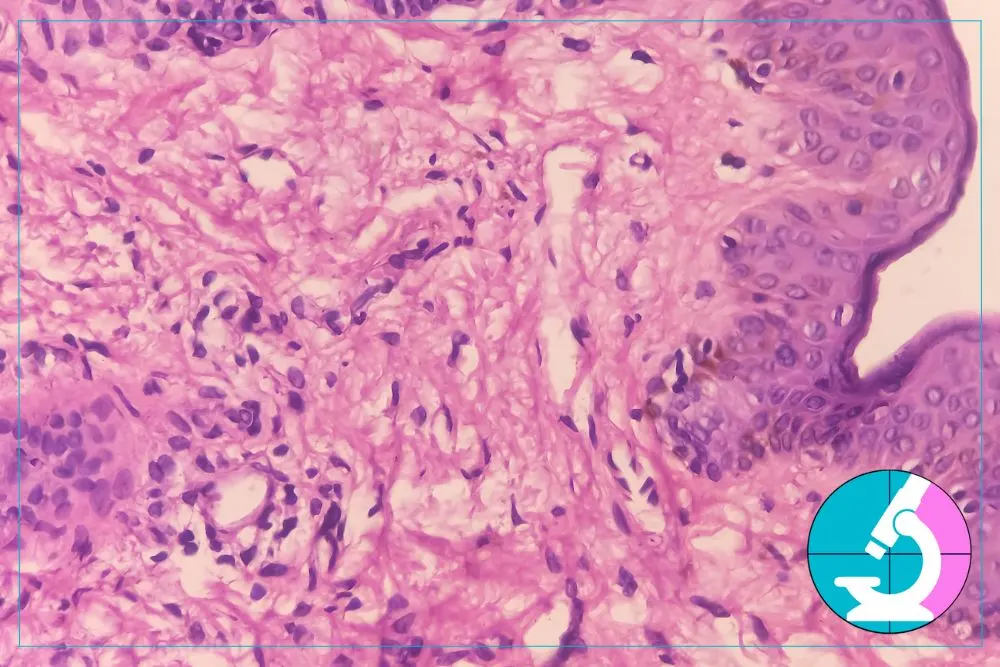
How a Cytopathologist Examines a Thyroid Biopsy
Once the sample has been prepared, the microscopic evaluation begins.
Cells collected during the biopsy are prepared on slides, stained, and examined under a microscope by a cytopathologist trained to spot subtle differences between benign and malignant cells.
This is where cytopathology becomes both science and experience.
I am looking at much more than whether cells appear “normal” or “abnormal.” Thyroid cells exhibit patterns, arrangements, nuclear characteristics, and other features that help us understand what process may be occurring within the nodule.
What I Look for Under the Microscope
During a Thyroid FNA evaluation, I may assess:
- Cellular arrangement
How thyroid cells group together can provide important information about the nature of a nodule. - Nuclear characteristics
The size, shape, appearance, and internal features of cell nuclei can help distinguish benign findings from suspicious ones. - Background material
Material surrounding the thyroid cells can also provide diagnostic clues. - The overall pattern
Cytopathology involves looking at the specimen as a whole rather than relying on a single abnormal looking cell.
Small differences can matter. This is why thyroid cytopathology requires specialized training and why the quality of both the sample and its interpretation are so important.
Why Rapid On Site Evaluation Matters During Fine Needle Aspiration
One of the most useful parts of my FNA process happens while the patient is still in the office.
Rapid On Site Evaluation, often shortened to ROSE, allows me to examine the sample immediately and determine whether I have collected enough diagnostic material.
Without on-site evaluation, a biopsy may be completed, and the patient may go home before the specimen is reviewed by a pathologist. If the sample later proves inadequate, the patient may need to return for another procedure.
I prefer to know while the patient is still sitting in front of me.
If the first sample does not contain enough cells, I can make another pass and collect additional material during the same visit.
This is especially valuable with thyroid nodules that are cystic, calcified, small, or otherwise challenging to sample.
ROSE does not mean I am rushing to make a final diagnosis during the procedure. It means I am making sure I have the material needed to perform a careful evaluation.
That distinction matters.
The goal is not simply to complete the biopsy. The goal is to obtain a useful biopsy.
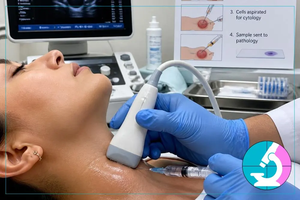
How Thyroid FNA Results Are Reached After the Sample Is Collected
After an adequate number of cells have been collected, the specimen undergoes a more detailed cytologic examination.
This is where the patterns seen under the microscope are considered together, and a diagnostic category is assigned.
Because Dr. Nadelman is both the FNA specialist and the cytopathologist reading the slides, patients avoid the delays and miscommunication that can happen when a biopsy and its interpretation are handled by two separate providers. You can learn more about this approach on my thyroid cancer and biopsy page.
Thyroid biopsy results are not always simply positive or negative for cancer. Cytopathologists use established terminology to communicate the degree of certainty and help referring physicians determine the appropriate next step.
A Thyroid Biopsy May Be Classified As
- Benign
The cells do not show evidence of malignancy, and the nodule can often be monitored rather than treated surgically. - Atypical or indeterminate
Some cellular features are unusual, but there is not enough evidence to confidently classify the nodule as benign or malignant. Repeat FNA or additional testing may be considered. - Suspicious
The cellular findings raise significant concern for cancer but may not meet every criterion needed for a definitive malignant diagnosis. - Malignant
The cytologic findings support a cancer diagnosis and help guide the patient’s next steps. - Non-diagnostic
There is not enough appropriate cellular material to provide a reliable interpretation. Immediate assessment of sample adequacy helps reduce the chance of this outcome.
A pathology report is ultimately a communication tool. It should help your physicians understand what was found and what should happen next.
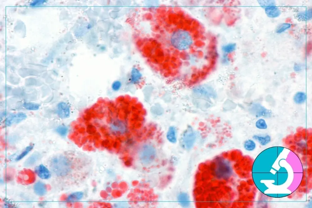
Why Having One FNA Thyroid Doctor Handle the Process Matters
In many healthcare systems, a thyroid biopsy involves several different people.
One physician may order the ultrasound. A radiologist may interpret it. Another physician may perform the biopsy. A laboratory processes the sample. Then a pathologist who has never met the patient reviews the slides.
That model can work, but it creates multiple handoffs.
My practice is intentionally different.
I review the ultrasound, perform the ultrasound guided Fine Needle Aspiration, evaluate the sample for adequacy, and interpret the cytology myself.
For me, those pieces belong together.
When I look at the cells under the microscope, I know where they came from because I was the physician who placed the needle. I know what the nodule looked like on ultrasound. I know which areas I sampled and whether there were any challenges during the procedure.
That context can be extremely useful when interpreting a thyroid biopsy.
It also makes communication simpler for the patient. Instead of wondering which office has the results or waiting for information to move between different providers, patients have one physician overseeing the diagnostic process.
Most patients receive a preliminary diagnosis within 24 hours, which helps reduce one of the hardest parts of any biopsy: waiting for answers.
What Your Thyroid Biopsy Sample Can Tell Us
It is remarkable how much information can come from such a small sample.
A few carefully collected cells can help determine whether a thyroid nodule can be safely monitored or requires additional evaluation.
For patients, the laboratory process can seem invisible. You see the ultrasound and experience the biopsy, but you do not see the careful slide preparation, microscopic examination, comparison of cellular patterns, and diagnostic reasoning that follow.
Those steps are every bit as important as the biopsy itself.
A good thyroid nodule biopsy requires three things to work together: precise sampling, proper specimen preparation, and experienced interpretation.
If any of those pieces is missing, the likelihood of an unclear result increases.
That is why I believe the person performing your Fine Needle Aspiration should understand cytopathology, and the person interpreting your cytology should understand exactly how the sample was obtained.
My goal is always to make that process as direct as possible for patients. You should understand why the biopsy is being performed, what happens to your sample, and what your results mean for your next step.
Contact an FNA Doctor About a Thyroid Nodule Biopsy
A thyroid biopsy can feel intimidating when you do not know what is happening behind the scenes. Once you understand the process, it becomes easier to see why each step matters.
From selecting the right area of a nodule to examining individual cells under the microscope, every part of the process contributes to the final diagnosis.
Patients travel to my Beverly Hills practice from throughout Southern California for thyroid Fine Needle Aspiration because they want a direct diagnostic experience with the same physician who performs and interprets their biopsy.
If you have a thyroid nodule that needs evaluation, you do not have to navigate that process without understanding what is happening.
Curious what your own thyroid FNA results could mean? Contact Dr. Nadelman to schedule a biopsy in Beverly Hills, with results in as little as 24 hours.
FAQs
Not always. Some samples are classified as atypical, indeterminate, suspicious, or non-diagnostic rather than clearly benign or malignant. These categories help physicians determine whether monitoring, repeat FNA, additional testing, or treatment should be considered.
Dr. Nadelman typically provides preliminary results within 24 hours. Because she performs the biopsy and interprets the cytology herself, the sample does not need to move between separate biopsy and pathology providers before preliminary findings can be communicated.
An inadequate sample can result in a non-diagnostic biopsy. Because Dr. Nadelman performs Rapid On Site Evaluation, she can assess specimen adequacy while the patient is still in the office and collect additional material during the same visit when necessary.
A cytopathologist is trained to recognize microscopic cellular patterns that help distinguish benign thyroid tissue from atypical, suspicious, or malignant findings. This microscopic examination provides information that ultrasound imaging alone cannot provide.
The collected cellular material is transferred to microscope slides and prepared so the cells can be examined by a cytopathologist. At Dr. Nadelman’s practice, the sample is also evaluated during the appointment to make sure adequate diagnostic material has been obtained.