
Why the Biopsy Method Matters
Finding a lump or a nodule on yourself can make every day of waiting feel long. The next step is not the same for every patient because different procedures answer different questions. Fine needle aspiration, core needle biopsy, and surgical biopsy vary in how much material they collect, what a pathologist can evaluate, and how invasive the procedure is. The right choice depends on the examination, imaging, whether the lump is solid or fluid filled, and which diagnosis must be confirmed or excluded.
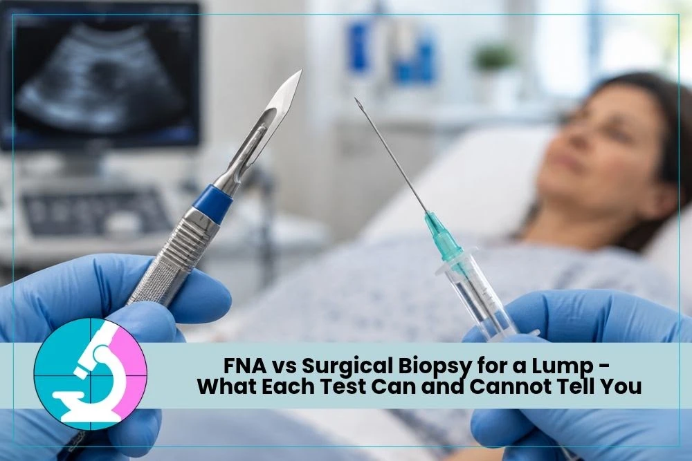
FNA removes cells through a very thin needle. A core biopsy removes narrow cylinders of tissue with a much larger needle. Surgical biopsy removes part or all of the abnormal area through a surgical incision. In current breast care, image-guided core biopsy is often the standard tissue test for a suspicious primary breast lesion. FNA still has valuable roles, especially when a rapid cytology answer can expedite care, when a cyst needs aspiration, or when a suspected metastasis in a lymph node is accessible. On the other hand, for thyroid nodules, a core biopsy is not the current standard of care, but rather an FNA is. Since the thyroid is a very vascular organ, anything larger than a fine needle biopsy can cause excessive bleeding.
The goal is to obtain enough information with the least risk and delay. Dr. Nadelman’s breast cancer and FNA resource and thyroid cancer and biopsy resource explain how cytology can contribute to the evaluation of breast and thyroid findings.
The type of sample gathered from an FNA is considered part of the realm of “cytology” and examines individual cells and small groups of cells (“cells in space”). An experienced cytopathologist looks at cell size, arrangement, background material, and other features in order to render a diagnosis. In contrast, the type of sample obtained from a core needle biopsy or excisional biopsy is considered a “surgical pathology” sample, where overall architecture and relationship of the cells to the surrounding tissue can be evaluated (e.g., invasion).
Standards are changing: Current practices are using smaller and smaller tissue samples
What FNA Can Show
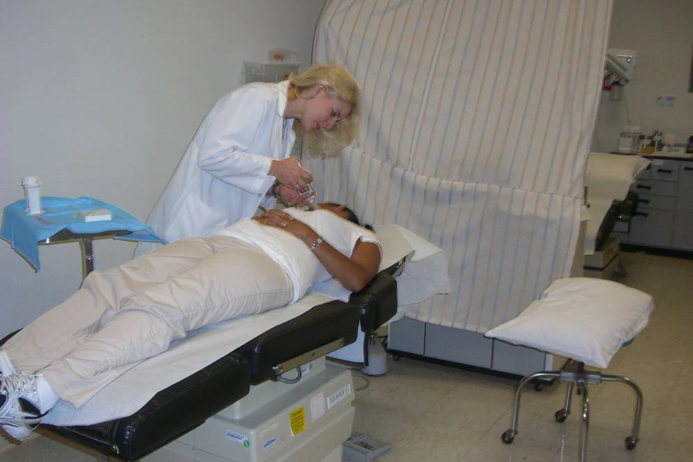
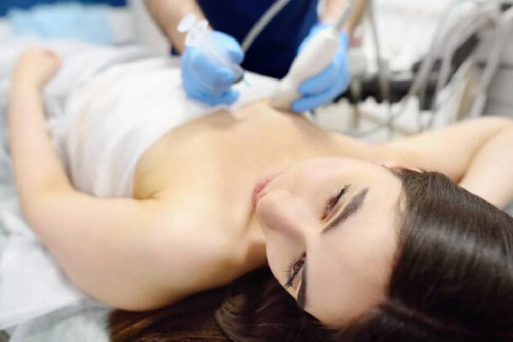
During a Fine Needle Aspiration biopsy, a physician guides a thin needle into the lump and draws out cells. Ultrasound guidance improves targeting and can help distinguish a solid mass from a cyst. The procedure is quick, usually leaves minor bruising, and does not require an incision or stitches. Rapid on-site evaluation, or ROSE, can confirm that the sample contains representative cells before the appointment ends. On site evaluation can help assess whether:
- Enough cells are present to eventually make a final diagnosis
- Sampled cells appear benign, atypical, suspicious, or malignant
- An enlarged lymph node contains metastatic cancer, lymphoma, infection-related changes, or another process
- Additional material is needed to create a cell block for ancillary testing such as immunostains, molecular tests, culture, flow cytometry, etc.
FNA can be especially useful when the clinical question is focused. For example, sampling a suspicious axillary lymph node may establish metastatic nodal involvement without surgery. FNA can also evaluate a palpable mass quickly and ultrasound-guided FNAs can evaluate nodules that are not felt. Dr. Nadelman’s FNA biopsy page describes how ultrasound guidance and immediate cytology review support an efficient visit.
What FNA May Not Be Able to Show
Because FNA collects cells rather than an intact tissue sample, it may not show how cells relate to surrounding tissue, such as ducts, stroma, or blood vessels. In a primary breast lesion, that can limit assessment of invasion. Distinguishing breast carcinoma in situ from invasive carcinoma generally requires tissue architecture.
FNA may also yield too few cells, particularly from small, fibrotic, calcified, cystic, or difficult-to-target lesions. A nondiagnostic result does not mean the lump is benign. It means the sample did not answer the question. The clinician must compare the results with the examination and imaging. If those findings are discordant, repeat sampling or a different biopsy method is necessary.
Breast cancer biomarkers such as ER, PR, and HER2 are commonly assessed on adequate tissue from core or surgical biopsy of the primary tumor. Some studies can be performed on cytology cell blocks in selected settings, but the team must know whether enough validated material is available. Treatment should not be planned from an incomplete sample when additional tissue is needed.
What Core and Surgical Biopsy Add
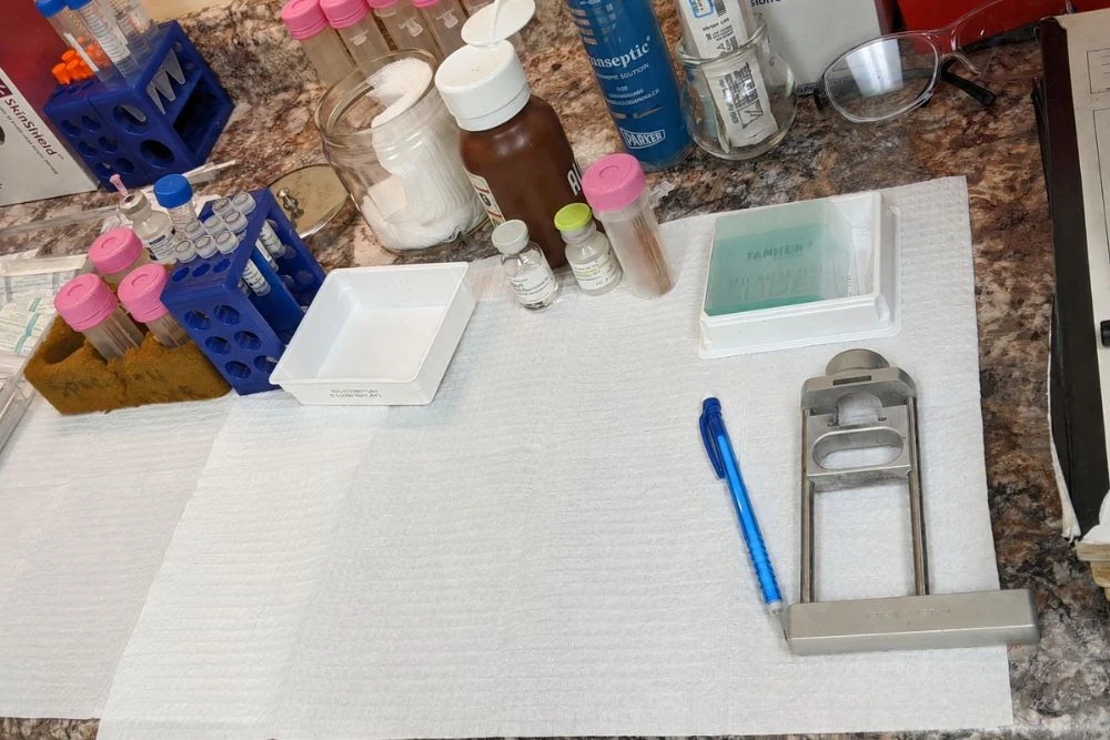
A core needle biopsy uses a spring-loaded or vacuum-assisted device to remove small tissue cylinders. Local anesthetic is used, and imaging guides the needle. The pathologist can assess cell features and tissue architecture, determine whether a carcinoma is invasive in many cases, and perform receptor testing on adequate tumor. Core biopsy usually provides the necessary diagnosis without an operation.
- Core biopsy is commonly chosen for a suspicious solid mass, architectural distortion, or imaging finding where invasion and biomarker testing may matter.
- Vacuum-assisted biopsy can collect additional tissue and is often used for certain calcifications or small imaging targets.
- Excisional surgical biopsy removes the entire abnormality and may be recommended when needle results are discordant, a high-risk lesion requires removal, or the target cannot be sampled reliably by needle.
- Incisional biopsy removes only part of a large abnormality and is used less often in routine breast diagnosis.
Surgical biopsy provides the largest specimen but carries greater burdens: an incision, scar, anesthesia, recovery, higher cost, and possible changes to later surgical planning. It should not be assumed to be the first or most accurate step in every case. Modern imaging-guided needle biopsy often gives the treatment team what it needs while preserving tissue.
Occasionally, surgery is the correct next step even after a needle biopsy. Examples include radiology-pathology discordance, a lesion that cannot be reached safely, or a result with uncertain malignant potential where complete excision is recommended. The recommendation should be explained in terms of the exact unanswered question.
The pathology report from a core biopsy may describe the tumor type, preliminary grade, and biomarkers, but the later surgical specimen can add information about final size, margins, and the relationship to nearby tissue and eventually staging. A patient should not be alarmed if the final report contains more detail or a refined classification. The two specimens serve different purposes, and some findings can only be measured after the lesion is removed.
When surgery is recommended, ask whether the procedure is diagnostic, therapeutic, or both. Also ask how the biopsy site will be marked, and whether additional imaging is needed. Thoughtful sequencing can reduce repeated procedures and preserve options. If a needle result is benign, confirm that the radiologist considers it concordant with the image rather than assuming the word benign ends the evaluation.
How Clinicians Choose the Right Test
The evaluation begins with a clinical exam and appropriate imaging, which may include diagnostic MRI, CT, mammography and ultrasound. The radiologist assigns a level of suspicion and identifies the safest route to the target. The clinician then chooses a sampling method that can answer the anticipated pathology question.
Your personal factors also matter. Blood-thinning medicines, bleeding disorders, prior surgery, ability to tolerate positioning, and urgency can affect the plan. Tell the team about all medicines and allergies. Ask about whether to stop a prescribed anticoagulant prior to the procedure.
A high-quality process includes radiology-pathology concordance. After the report is issued, the radiologist and treating clinician decide whether the result explains the imaging finding. A benign result that fits a benign-appearing lesion may lead to routine or short-interval follow-up. A benign result that does not fit a suspicious image requires more evaluation.
Frequently Asked Questions
Is FNA less accurate than core biopsy?
Accuracy depends on the target, the question, sampling quality, and pathology expertise. FNA can be highly accurate for selected palpable lesions and lymph nodes, especially with ultrasound guidance and ROSE. Core biopsy is preferred when tissue architecture, invasion, or receptor testing is required.
Can FNA tell whether a breast cancer is invasive?
FNA cytology can identify malignant cells but usually cannot reliably determine whether a primary breast carcinoma is in situ or invasive because it does not preserve enough surrounding architecture. Core or surgical tissue is generally needed to assess invasion.
Will a needle biopsy spread breast cancer?
Needle biopsy is a standard diagnostic procedure, and clinically meaningful tumor spread along the needle path is exceedingly rare, if at all. The benefit of obtaining an accurate diagnosis and treatment plan greatly outweighs that theoretical concern for appropriately selected patients.
What happens after a nondiagnostic FNA?
The team reviews whether the needle reached the correct target and whether the imaging remains suspicious. Options include repeat FNA with ROSE, core biopsy, vacuum-assisted biopsy, surgical excision, or imaging follow-up when the overall findings support it. A nondiagnostic sample should not be treated as a benign diagnosis.
How long does recovery take?
After FNA, most patients resume light activity immediately and may have bruising and mild soreness. Core biopsy may cause more bruising and usually requires a dressing and short activity limits. Surgical biopsy has a longer recovery that depends on the incision, anesthesia, and procedure details.
Choose the Test That Answers the Clinical Question
A breast lump deserves a clear plan rather than a reflexive choice between the smallest and largest procedure. FNA can offer fast, minimally invasive cytology. Core biopsy supplies architecture and is often best for evaluating invasion and biomarkers. Surgical biopsy is reserved for situations in which needle methods cannot provide a concordant answer or removal is itself recommended.
Not sure which biopsy is right for a breast lump? Contact Dr. Nadelman to discuss whether FNA, core biopsy, or another diagnostic step is most appropriate.