
Understanding Papillary vs. Follicular Thyroid Cancer
Hearing the words “thyroid cancer” can be frightening, but thyroid cancer is not one single disease. There are several types, and understanding which type may be present is important because they can behave, spread, and be diagnosed differently.
Papillary and follicular thyroid cancer are the two most common types of differentiated thyroid cancer. Both begin in thyroid follicular cells, the cells responsible for producing and storing thyroid hormones. Both also tend to grow relatively slowly and are often highly treatable.
That does not mean they are identical.
Papillary thyroid cancer has distinctive cellular features that can often be recognized through Fine Needle Aspiration. Follicular thyroid lesions present a different diagnostic challenge because distinguishing a benign follicular adenoma from follicular carcinoma requires determining whether the tumor has invaded its capsule or blood vessels.
That distinction cannot be made from an FNA sample alone.
Papillary and follicular thyroid cancer are the two most common types of differentiated thyroid cancer, and evaluating a suspicious nodule starts with an accurate biopsy. Patients can learn more about thyroid cancer and biopsy and how these diagnoses are approached.
As a cytopathologist and Fine Needle Aspiration specialist, my role is to obtain the best possible cellular sample, interpret what those cells tell us, and recognize when the findings require another diagnostic step.
How Papillary Thyroid Cancer Differs From Follicular Thyroid Cancer
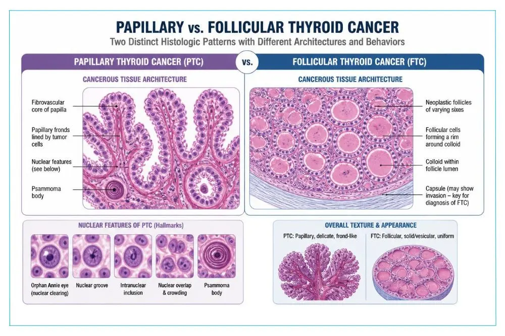
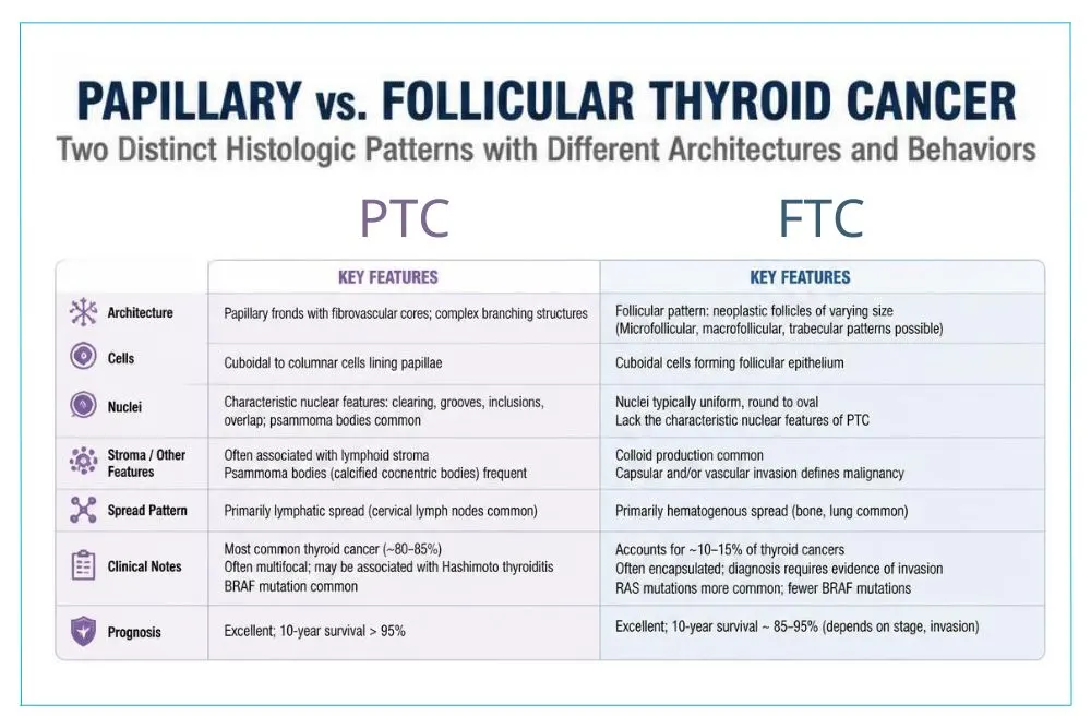
Papillary and follicular thyroid cancers share an origin in thyroid follicular cells, but their microscopic appearance and patterns of spread are different.
Papillary thyroid carcinoma is the more common of the two. One of the advantages we have as cytopathologists is that papillary thyroid cancer often produces recognizable cellular changes that can be seen in a properly collected FNA specimen.
Follicular lesions are more complicated.
Key Differences Between Papillary and Follicular Thyroid Cancer
- Papillary thyroid cancer has characteristic cellular features
Cytopathologists can often identify the nuclear changes associated with papillary thyroid carcinoma in an FNA sample, allowing a diagnosis without first removing the entire nodule. - Follicular thyroid cancer requires evaluation of invasion
The cells of a follicular adenoma and follicular carcinoma can look very similar. To diagnose carcinoma, a pathologist needs to determine whether the tumor has invaded through its capsule or into blood vessels. - The cancers can spread differently
Papillary thyroid carcinoma tends to spread through lymphatic channels and may involve lymph nodes in the neck. Follicular thyroid carcinoma is more likely to spread through the bloodstream when it metastasizes. - Both are differentiated thyroid cancers
This means the cancer cells retain some characteristics of normal thyroid cells. In general, differentiated thyroid cancers are associated with favorable outcomes, particularly when identified and treated appropriately.
These differences are important because a thyroid biopsy is not simply about answering “cancer or no cancer.” It is about understanding what the cells can tell us and, equally important, recognizing what they cannot.
How Fine Needle Aspiration Helps Diagnose Papillary Thyroid Cancer
Fine Needle Aspiration is the standard first-line biopsy method for evaluating suspicious thyroid nodules.
During the procedure, I use ultrasound guidance to place a very thin needle directly into the area of the nodule I want to evaluate. Cells are collected and examined under the microscope.
Both papillary and follicular thyroid lesions can be evaluated with FNA, but the information we obtain from each differs.
Papillary thyroid carcinoma often has characteristic nuclear features that a cytopathologist can recognize. These microscopic changes can make Fine Needle Aspiration extremely useful in diagnosing papillary thyroid cancer.
What I Evaluate During a Thyroid FNA
- Cellular architecture
How the thyroid cells arrange themselves can provide important clues about the type of lesion being sampled. - Nuclear characteristics
Papillary thyroid carcinoma is associated with distinctive nuclear changes. Recognizing these subtle features requires experience in thyroid cytopathology. - Background material and other cells
The material surrounding the thyroid cells can provide additional diagnostic information and may help place the cellular findings in context. - Sample adequacy
I use Rapid On Site Evaluation to make sure I have collected enough useful material before the patient leaves my office.
Fine Needle Aspiration is minimally invasive and provides important diagnostic information without requiring surgery. My FNA biopsy page explains how the procedure works and what patients can expect.
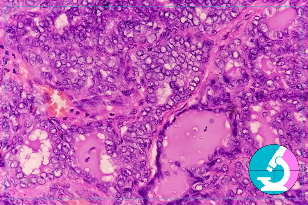
Why Follicular Thyroid Cancer Is Different on a Thyroid Biopsy
Follicular thyroid lesions are one of the best examples of why understanding the limitations of a biopsy is just as important as understanding its strengths.
An FNA can identify a follicular pattern and help determine whether a nodule requires further evaluation. What it cannot do is distinguish with certainty between a follicular adenoma and a follicular carcinoma.
Why?
The difference is not simply how the individual cells look.
A follicular adenoma is a benign tumor surrounded by a capsule. Follicular carcinoma is diagnosed when tumor cells invade that capsule or enter blood vessels. To see that invasion, a pathologist must examine the relationship between the tumor and its entire capsule after the nodule has been surgically removed.
A Fine Needle Aspiration samples cells from within the nodule. It does not remove the capsule.
Importantly, a core needle biopsy does not solve this problem either. A core biopsy only removes a portion of the lesion and still cannot demonstrate the status of the entire capsule. If a follicular neoplasm requires definitive distinction between adenoma and carcinoma, surgical excision is necessary to allow thorough examination of the capsule.
This is why the wording on a thyroid pathology report matters so much. A follicular pattern on FNA should not be presented to a patient as a definitive diagnosis of follicular carcinoma when the required evidence of invasion cannot yet be evaluated.
An accurate diagnosis sometimes means knowing when the cells have given us the answer and when another step is necessary.
How Papillary and Follicular Thyroid Cancer Can Behave Differently
Both papillary and follicular thyroid cancer tend to be slow-growing, and both are often highly treatable. However, their different patterns of spread can influence how physicians evaluate and monitor patients.
Differences Physicians Consider
- Lymph node involvement
Papillary thyroid carcinoma commonly spreads through the lymphatic system, which is why the lymph nodes of the neck are carefully evaluated when papillary cancer is suspected or diagnosed. - Bloodstream spread
Follicular thyroid carcinoma is more likely than papillary carcinoma to spread through blood vessels. When metastatic disease occurs, distant sites such as the lungs or bones may require evaluation. - Nodule characteristics
Ultrasound findings provide valuable information about a thyroid nodule, but imaging must be considered alongside cytopathology and the patient’s clinical history. - Individual patient factors
Age, medical history, nodule characteristics, symptoms, and other findings all contribute to decisions about treatment and follow-up.
These differences are one reason I believe thyroid nodule evaluation needs to remain individualized. A diagnosis should never be reduced to a single ultrasound score, pathology phrase, or isolated finding.

Why Your FNA Doctor and Cytopathologist Matter
Patients sometimes think of a thyroid biopsy as a mechanical procedure: put a needle into a nodule, send the sample to a laboratory, and wait for the answer.
There is much more to it.
The quality of the ultrasound evaluation affects where the needle is placed. The quality of the sampling affects which cells are available for review. The preparation of those cells affects what can be seen under the microscope. Finally, the cytopathologist’s experience affects how those findings are interpreted.
In many healthcare settings, those responsibilities are divided among several people.
My practice is different because I perform the ultrasound guided FNA and interpret the cytology myself. That allows me to connect what I saw on ultrasound with what I see under the microscope.
It also allows me to perform Rapid On Site Evaluation during the procedure. If I need additional cells, I know while the patient is still with me rather than discovering later that another biopsy may be necessary.
For papillary thyroid carcinoma, careful cytologic evaluation can provide a highly informative diagnosis. For a follicular lesion, expertise is equally important because the correct interpretation may be recognizing that the cells alone cannot answer the adenoma versus carcinoma question.
Both types tend to be slow-growing and highly treatable, but they can behave differently and require different monitoring, which is why a precise FNA reading matters.
My goal is to give patients as much clarity as the specimen can responsibly provide without overstating what a biopsy can tell us.
Contact an FNA Thyroid Doctor About Papillary vs. Follicular Thyroid Cancer
If you have been told you have a suspicious thyroid nodule, an abnormal ultrasound, or an unclear biopsy result, it is understandable to want answers quickly.
Most thyroid nodules are benign. Even when thyroid cancer is diagnosed, papillary and follicular thyroid cancers are often slow-growing and highly treatable. Getting an accurate diagnosis is what allows you and your physicians to decide what should happen next without making assumptions.
Dr. Nadelman’s dual role as an FNA specialist and cytopathologist means the same physician who performs your biopsy also interprets the cells, helping to provide continuity throughout the diagnostic process.
Patients travel to my Beverly Hills practice from throughout Southern California for thyroid Fine Needle Aspiration and cytopathology evaluation. Next day appointments are often available, and preliminary FNA results are typically available within 24 hours.
Concerned about a thyroid nodule? Contact Dr. Nadelman for a consultation in Beverly Hills, with next-day appointments and house calls available throughout California.
FAQs
Both are often highly treatable, particularly when diagnosed and managed appropriately. Treatment depends on the specific diagnosis, extent of disease, individual patient factors, and other clinical findings.
No. A follicular pattern on an FNA biopsy does not automatically mean follicular carcinoma. Benign follicular adenomas can have similar cytologic appearances, which is why additional evaluation may be required before a definitive diagnosis can be made.
No. FNA can identify a follicular pattern or follicular neoplasm, but distinguishing a benign follicular adenoma from follicular carcinoma requires evaluation for capsular or vascular invasion. That requires examination of the surgically excised nodule and its capsule.
Fine Needle Aspiration can often identify papillary thyroid carcinoma because the cancer has characteristic cellular and nuclear features that an experienced cytopathologist can recognize. Ultrasound guided sampling and adequate cellular material are important for an accurate evaluation.
Yes. Papillary thyroid carcinoma is the most common type of thyroid cancer. Follicular thyroid carcinoma is less common, although both originate from thyroid follicular cells and are considered differentiated thyroid cancers.