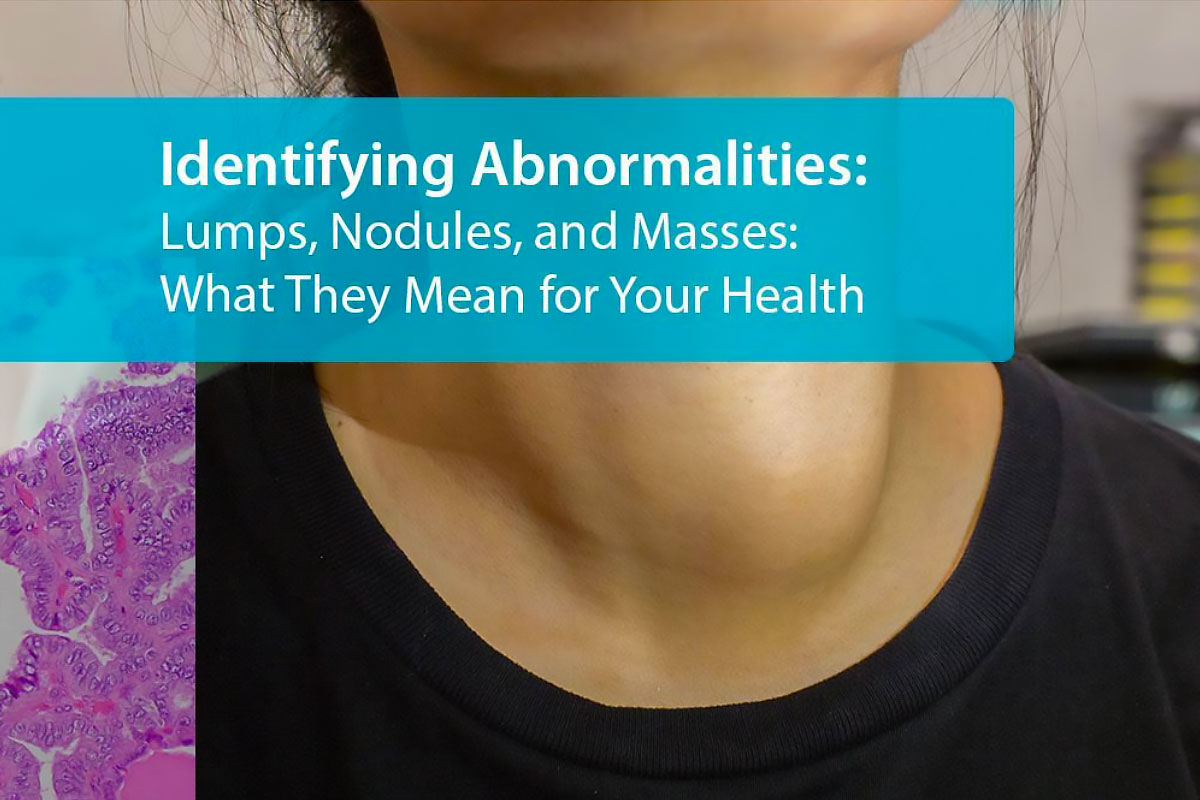
Detecting lumps under skin and other physical abnormalities early is crucial for effective health monitoring and can be pivotal in diagnosing various conditions, ranging from benign lumps to potentially cancerous lumps and life-threatening diseases. An unexpected lump, nodule, or mass under the skin often serves as the first tangible sign that prompts individuals to seek medical attention. For instance, consider Sarah, who noticed a small, painless lump in her breast during a routine self-examination. Initially hesitant, Sarah’s decision to promptly consult her healthcare provider led to an early diagnosis of a benign growth, alleviating her fears and allowing for timely management. Sarah’s story underscores the commonality of such discoveries and highlights the critical importance of not dismissing lumps and bumps in our bodies, no matter how insignificant they may seem.

Deciphering Lumps, Nodules, and Masses
The terms “lumps,” “nodules,” and “masses” often evoke concern, but understanding their definitions and distinctions can provide clarity and ease anxieties. Lumps are defined as any noticeable bump or swelling under the skin, varying widely in size and consistency. Nodules are a subtype of lumps, usually described as small, often solid, raised areas deeper within or just under the skin. Masses are generally larger formations that can be benign (non-cancerous lumps) or malignant (cancerous lumps), necessitating further medical evaluation to determine their nature.
Despite their differences, these terms are sometimes used interchangeably in casual discussion. However, medical professionals distinguish between them based on specific criteria, including size, location, and physical characteristics, to guide diagnosis and treatment using imaging such as CT scan or MRI scan for accuracy.
Anatomical Occurrences
Lumps, nodules, and masses can occur in various parts of the body, each presenting unique considerations based on their location:
- Breast: Discoveries in the breast, such as lumps or masses, are common concerns leading to medical consultations. While often benign lumps, such as fibrocystic changes or cysts, they require evaluation to rule out breast cancer.
- Thyroid: Thyroid nodules in the thyroid gland are common and usually benign. However, they can occasionally indicate thyroid cancer or cause hormonal imbalances involving thyroid hormones, affecting overall health.
- Lymph Nodes: Swollen lymph nodes, particularly those that are firm or persistently enlarged, can be signs of infection or, less commonly, lymphoma or other cancers.
- Skin: Skin lumps can range from benign ganglion cysts and lipomas to malignant melanomas. The skin’s visibility makes it easier to notice changes, emphasizing the importance of regular self-examination.
The significance of these locations lies in their association with different types of tissues and potential health implications. For example, lumps in the breast or thyroid may prompt concerns about cancer, whereas skin lumps could vary widely from harmless cysts to urgent melanomas. Understanding these anatomical occurrences is vital for interpreting the potential health implications of discovered abnormalities and underscores the necessity of medical evaluation for accurate diagnosis and appropriate management.

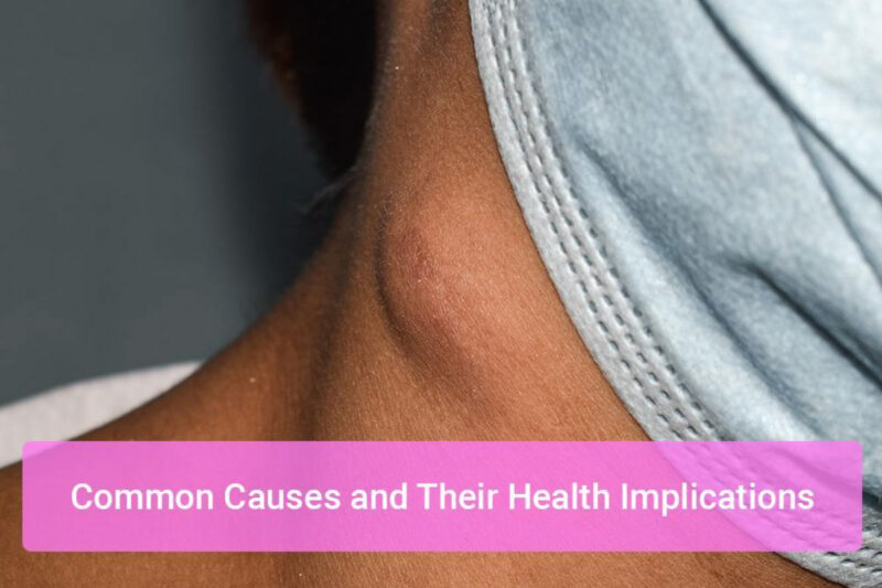
Common Causes For Lumps and Their Health Implications
When an individual discovers a lump, nodule, or mass, understanding whether it is benign or malignant is crucial. Here we explore the differences between these conditions, their causes, and health implications.
Benign vs. Malignant Lump
Benign growths are non-cancerous and generally do not spread to other parts of the body. They can grow but typically do so at a slower rate and are less likely to recur after removal. Malignant growths, on the other hand, are cancerous lumps. They have the potential to invade nearby tissues and spread to other body parts, a process known as metastasis.
While the exact probability of a lump being malignant varies by factors such as age, family history, and the lump’s characteristics, statistical insights show that the majority of breast lumps are benign lumps, particularly in women under 40. However, the risk of malignancy increases with age, making early detection, screening, and CT scans or MRI scans paramount.
Cysts
- Types: Sebaceous cysts form in skin glands, while ovarian cysts develop in the ovaries.
- Causes: Blocked glands, hormonal imbalances, or infections can lead to cyst formation.
- Symptoms: Swelling, pain if infected, or no symptoms at all.
- Treatment: Many cysts resolve without treatment, but persistent or symptomatic cysts may require drainage or surgical removal.
Cysts are a common type of lumps under the skin, and while most lumps are harmless, some may resemble ganglion cysts or other benign growths that can cause discomfort.
Benign Tumors
- Examples: Fibroadenomas are common in the breast, while lipomas, composed of fat cells, can appear anywhere in the body.
- Growth and Discomfort: They can grow and sometimes cause discomfort or interfere with normal body functions.
- Treatment: Observation is often recommended for small, asymptomatic tumors. Surgical removal may be necessary if the tumor causes symptoms or grows.
Most benign lumps or lumps and bumps are not a cause for alarm, but if you are worried about a lump, especially one that changes in size or causes pain, medical evaluation is important.
Infections and Abscesses
- Formation: Bacterial infections can lead to the accumulation of pus in tissue, forming an abscess.
- Immune System Role: The body’s immune response to infection can cause inflammation and swelling.
- Treatment: Antibiotics for the infection and drainage of the abscess are common treatments.
Infections can sometimes mimic lumps under the skin, leading to swelling that may appear similar to a cyst or benign lump.
Inflammatory Conditions
- Conditions: Sarcoidosis can cause granulomas, while autoimmune diseases like rheumatoid arthritis might lead to nodules.
- Treatment: Managing inflammation and the underlying condition is key, often involving corticosteroids or other immunosuppressive medications.
In some inflammatory conditions, lung nodules can appear on imaging such as a CT scan, sometimes accompanied by shortness of breath. Other cases, like thyroid disorders, may show thyroid nodules that influence thyroid hormones.
Cancer
- Common Cancers: Breast, thyroid, and skin cancers (melanoma) are among the types most likely to present with palpable lumps.
- Screening and Detection: Regular screenings, such as mammograms for breast cancer, are vital for early detection. The earlier cancerous lumps are found, the more effective treatment is likely to be.
Understanding the nature of lumps, nodules, and masses is essential for appropriate management and treatment. While the discovery of such abnormalities can be alarming, it’s important to remember that many are benign. Prompt medical evaluation is key to determining the cause and deciding on the best course of action. Early detection and treatment are crucial, especially for malignant growths, underscoring the importance of regular health screenings and self-examinations.
What to Do If You Discover a Lump or Abnormality
Discovering an abnormality on your body can be unsettling. However, taking a methodical approach can help you provide valuable information to your healthcare provider, leading to a more accurate diagnosis. Here’s what to do if you find a lump, nodule, or mass.
Initial Steps
- Measure the Abnormality: Using a flexible tape measure, note the size of the lump. If it’s not easily measurable, compare it to a common object (e.g., a pea or a marble).
- Document Characteristics: Write down the lump’s characteristics – is it hard or soft? Fixed in place or movable? Painful to touch or not?
- Monitor Changes: Keep a log of any changes in the lump or nodule over time. This includes size, texture, and any new symptoms like pain, redness, or shortness of breath. Noting these changes can be crucial for your doctor’s assessment.
- Photographic Documentation: If visible, take photographs from different angles to track visible changes over time. Ensure consistent lighting and distance for accuracy when observing lumps and bumps.
Seeking Professional Help
- Gather Your Medical History: Compile a detailed medical history, including any previous illnesses, surgeries, medications, and any family history of similar conditions or benign growths or cancerous lumps.
- Prepare a List of Questions: Write down any questions or concerns you have about the lump or nodule to ensure you do not forget to address them during your visit.
Questions might include
- What potential causes are there for this type of lump?
- What tests or examinations will we use to investigate further, such as CT scans, MRI scans, or ultrasound imaging?
- Based on my medical history, how concerned should we be about cancerous lumps or benign tumors?
- What to Bring: In addition to your medical history and list of questions, bring any relevant insurance information and a notebook or electronic device to take notes during your appointment.
If you’re worried about a lump, don’t delay getting a professional opinion. Some lumps are harmless, but early evaluation prevents complications.
Diagnostic Process for Lumps
Your doctor will first perform a physical examination of the lump, noting its size, texture, and location. Depending on the type of lumps and bumps, your doctor may recommend imaging tests such as:
- Mammography: Especially for breast lumps, to look for signs of cancer.
- Ultrasound: Useful for distinguishing between solid and fluid-filled lumps under the skin.
- CT or MRI Scans: These can provide detailed images of the soft tissue lump and surrounding areas, helping to identify the lump’s nature and whether it has affected nearby structures.
- Biopsy: If imaging tests suggest the lump could be cancerous, or if its nature remains unclear, a biopsy might be performed. This involves removing a small tissue sample from the lump for laboratory analysis. Biopsies can be done using a fine needle aspiration, a larger needle (core biopsy), or through a minor surgical procedure.
- Further Tests: Based on the biopsy results, additional tests may be recommended to determine the lump’s cause and whether it has spread.
Discovering an abnormality can be the first step in addressing potential health issues. By measuring, documenting, and seeking professional help, you can ensure that you receive a thorough evaluation and appropriate care. Early detection and intervention are key to managing health concerns effectively.
Prevention and Early Detection of Lumps
While not all lumps, nodules, and masses can be prevented, adopting a proactive approach to your health can aid in early detection. Regular self-examinations and participation in health screening programs for conditions like breast cancer, thyroid nodules, or lung nodules are effective strategies.
Additionally, maintaining a healthy lifestyle, including a balanced diet and regular exercise, can help reduce the risk of certain types of cancers and inflammatory conditions. Understanding when to worry about a lump and getting it checked early can save lives.

The Role of Healthcare Providers in Managing Lumps and Abnormalities
Upon finding an abnormality, healthcare providers play a crucial role in diagnosis and treatment. They will consider factors such as your medical history, the abnormality’s characteristics, and any associated symptoms to develop a management plan. This plan may include monitoring the lump over time, prescribing medication, or in some cases, performing surgery.
Your provider may also order a CT scan, MRI scan, or thyroid hormone evaluation depending on your symptoms and the location of the lump.
Conclusion
Discovering a lump, nodule, or mass can be a source of anxiety, but understanding the potential causes and appropriate steps to take can empower you to manage your health effectively. Many lumps under the skin are benign lumps or ganglion cysts that don’t pose serious risks.
However, cancerous lumps require early attention. Always consult with a healthcare provider for an accurate diagnosis and personalized advice. Remember, early detection, fine needle aspiration, and regular medical evaluation play vital roles in ensuring your long-term health and well-being.

References
Daly, C., & Puckett, Y. (2022). New Breast Mass. In StatPearls. StatPearls Publishing.
Expert Panel on Breast Imaging:, Moy, L., Heller, S. L., Bailey, L., D’Orsi, C., DiFlorio, R. M., Green, E. D., Holbrook, A. I., Lee, S. J., Lourenco, A. P., Mainiero, M. B., Sepulveda, K. A., Slanetz, P. J., Trikha, S., Yepes, M. M., & Newell, M. S. (2017). ACR Appropriateness Criteria® Palpable Breast Masses. Journal of the American College of Radiology : JACR, 14(5S), S203–S224. https://doi.org/10.1016/j.jacr.2017.02.033
Expert Panel on Breast Imaging:, Moy, L., Heller, S. L., Bailey, L., D’Orsi, C., DiFlorio, R. M., Green, E. D., Holbrook, A. I., Lee, S. J., Lourenco, A. P., Mainiero, M. B., Sepulveda, K. A., Slanetz, P. J., Trikha, S., Yepes, M. M., & Newell, M. S. (2017). ACR Appropriateness Criteria® Palpable Breast Masses. Journal of the American College of Radiology : JACR, 14(5S), S203–S224. https://doi.org/10.1016/j.jacr.2017.02.033
Expert Panel on Breast Imaging:, Lee, S. J., Trikha, S., Moy, L., Baron, P., diFlorio, R. M., Green, E. D., Heller, S. L., Holbrook, A. I., Lewin, A. A., Lourenco, A. P., Niell, B. L., Slanetz, P. J., Stuckey, A. R., Vincoff, N. S., Weinstein, S. P., Yepes, M. M., & Newell, M. S. (2017). ACR Appropriateness Criteria® Evaluation of Nipple Discharge. Journal of the American College of Radiology : JACR, 14(5S), S138–S153. https://doi.org/10.1016/j.jacr.2017.01.030
Expert Panel on Breast Imaging:, Jokich, P. M., Bailey, L., D’Orsi, C., Green, E. D., Holbrook, A. I., Lee, S. J., Lourenco, A. P., Mainiero, M. B., Moy, L., Sepulveda, K. A., Slanetz, P. J., Trikha, S., Yepes, M. M., & Newell, M. S. (2017). ACR Appropriateness Criteria® Breast Pain. Journal of the American College of Radiology : JACR, 14(5S), S25–S33. https://doi.org/10.1016/j.jacr.2017.01.028
Klein S. (2005). Evaluation of palpable breast masses. American family physician, 71(9), 1731–1738.
Chung, H. L., Le-Petross, H. T., & Leung, J. W. T. (2021). Imaging Updates to Breast Cancer Lymph Node Management. Radiographics : a review publication of the Radiological Society of North America, Inc, 41(5), 1283–1299. https://doi.org/10.1148/rg.2021210053
Balla, A., & Weaver, D. L. (2022). Pathologic Evaluation of Lymph Nodes in Breast Cancer: Contemporary Approaches and Clinical Implications. Surgical pathology clinics, 15(1), 15–27. https://doi.org/10.1016/j.path.2021.11.002


