

The discovery of a new growth or mass can be filled with uncertainty and concern. It often begins with a personal discovery, a routine clinical assessment, or a revealing imaging scan. The journey from discovery to diagnosis is a critical path, marked by the need for swift and precise answers. In modern medicine, where the stakes are high, and the need for accuracy is paramount, Fine Needle Aspiration (FNA) has emerged as an indispensable tool. Its role extends beyond mere diagnosis; it’s an integral part of the sophisticated puzzle of cancer treatment planning, especially in the era of personalized medicine and targeted therapies.
The Discovery of New Growth or Mass

The discovery of a new growth or mass, whether through personal examination, clinical assessment, or imaging, can be a significant concern for patients. Swift and accurate diagnosis, minimizing psychological and physical stress for the patient, is a paramount goal of healthcare teams. While history-taking, physical examination, and advanced imaging can diagnose many new masses confidently, certain cases may present diagnostic uncertainty.
Obtaining a tissue or fluid sample through FNA is essential in such scenarios. Moreover, in the context of advanced biological drugs and genotyping, FNA coupled with genetic and molecular testing becomes indispensable in formulating the most effective treatment plan.
Fine Needle Aspiration: An Indispensable Tool in Oncological Diagnostics
Fine Needle Aspiration (FNA) biopsy, a cornerstone in cancer diagnostics, is a minimally invasive procedure involving a thin, hollow needle to extract cellular samples from suspicious lesions. This technique is particularly pivotal in the diagnosis of breast and thyroid cancer, where early and accurate detection is crucial for effective treatment planning.
The Role of Ultrasound and Imaging in FNA
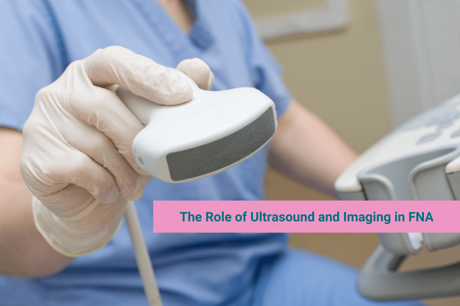
Ultrasound-guided FNA biopsies have enhanced the precision of the procedure, allowing real-time visualization of the needle path for accurate lesion targeting. This advancement has significantly reduced the risk of complications and improved diagnostic accuracy.
FNA is versatile and applicable across various body regions, including breast, thyroid, lymph nodes, and suspicious skin masses. The advent of endoscopic ultrasound has further expanded its use to include biopsies of pancreatic, gastrointestinal, esophageal, and tracheal pathologies. With the advancement of imaging technologies, CT-guided FNA can target almost any body region. Key to successful FNA is careful planning to avoid critical structures and minimize risks, particularly in infected areas or regions with complex anatomy.
The Science Behind FNA Biopsy in Cancer Detection
FNA biopsy’s utility lies in its ability to provide rapid cytological evaluation. When a patient presents with a breast lesion, the FNA biopsy can ascertain the presence of malignant cells. This procedure is less invasive than traditional surgical biopsies and can be performed with minimal discomfort to the patient.
Comparing FNA with Core Needle Biopsy: A Deeper Insight
In breast cancer diagnosis, FNA is often weighed against core needle biopsy. The latter, slightly more invasive, provides larger tissue samples for histopathology, essential for identifying specific breast cancer subtypes. This distinction is critical in tailoring treatment strategies, such as the choice between hormone therapy, immunotherapy, and chemotherapy.
Sensitivity and specificity are crucial metrics when evaluating the efficacy of Fine Needle Aspiration Cytology (FNAC) and Core Needle Biopsy (CNB). These metrics directly influence the predictive values for both negative and positive diagnoses.
Studies have shown varying results, but overall, CNB tends to exhibit higher sensitivity and specificity compared to FNAC. This is particularly notable in diagnosing benign and malignant breast lesions.
For example, one large study reported high sensitivity (97.1%) and specificity (99.1%) for FNAC. However, it’s important to note that this study excluded atypical and suspicious categories, which can represent a significant portion of breast lesions.
The performance of FNAC in diagnosing malignancy is generally comparable to CNB, especially in definitive lesions. However, for suspicious or atypical lesions, CNB often achieves higher performance indices. The positive predictive value for FNAC drops significantly for suspicious lesions and atypia, whereas CNB maintains higher rates in these categories.
Advantages and Disadvantages of FNA
While methodologically different, FNAC and CNB have unique advantages and limitations. FNAC is more suitable for patients on anticoagulants, lesions near critical areas (such as the skin, chest wall, vessels, or implants), and small or deep-seated lesions. For accessible, palpable lesions, FNAC is relatively straightforward and quick, making it a convenient choice in outpatient settings.
However, the quality of FNAC largely depends on the expertise of the individual performing the aspiration and the (cyto)pathologist interpreting the results. This dependency on skill and experience is critical in choosing between FNAC and CNB.
FNA in Comprehensive Cancer Care: From Diagnosis to Treatment Monitoring
Fine-needle aspiration (FNA) is a versatile and minimally invasive diagnostic tool used in various clinical scenarios. It is particularly effective in evaluating thyroid nodules, skin masses, breast lumps, and suspicious lymph nodes for potential malignancy. FNA is instrumental in initial diagnosis and plays a crucial role in cancer staging, monitoring treatment efficacy, and detecting recurrences. This technique is invaluable in the ongoing management of known metastatic diseases, where it assists in identifying appropriate chemotherapeutic or biological treatments based on genetic or molecular markers. Additionally, FNA has therapeutic applications, such as aspirating the contents of abscesses in cosmetically sensitive areas like the breast, offering an alternative to more invasive procedures like incision and drainage. Its less invasive nature makes FNA an ideal choice for repeated tissue sampling, a critical aspect in the dynamic field of cancer treatment where ongoing assessment of patient response is essential.
Sample Analysis and Hormone Receptor Detection
Post-biopsy analysis is a critical step in personalizing cancer treatment, particularly in breast cancer. After a biopsy, such as fine-needle aspiration, the collected tissue undergoes various tests, including estrogen (ER) and progesterone receptor (PR) tests. These tests are crucial in determining the presence of hormone receptors in the cancer cells, which directly influences the suitability and effectiveness of hormone therapy. Hormone therapies target these receptors to slow down or stop the growth of hormone-sensitive tumors.
In cases where breast cancer cells are found to be HER2-positive, identified through specific tests conducted on the biopsy samples, targeted therapies play a central role. HER2-positive breast cancers are known to be more aggressive, and the overexpression of the HER2 protein leads to rapid cell growth and division. In this case, targeted therapies are designed to specifically attack the HER2 protein, thereby slowing the growth and spread of the cancer.
Moreover, understanding the hormonal and HER2 status of breast cancer allows oncologists to tailor the treatment plan more precisely. This approach not only improves the treatment’s effectiveness but also minimizes patients’ exposure to potentially unnecessary or less effective treatments. Therefore, post-biopsy analysis, including hormone receptor and HER2 status testing, is vital for guiding treatment decisions and improving patient outcomes in breast cancer care.
Safety and Efficacy: The Clinical Profile of FNA Biopsy
The safety profile of FNA biopsies is favorable, with a low risk of complications such as bleeding or infection. The procedure’s minimal invasiveness also facilitates faster recovery times, making it a preferred choice for patients and clinicians. Seek care from Dr. Celina M. Nadelman in Beverly Hills, Los Angeles, California, for your FNA biopsy.
What should you do to prepare for FNA?
Preparing for a fine needle aspiration biopsy is relatively straightforward. You may need to temporarily stop taking certain medications, especially blood thinners like aspirin, clopidogrel (Plavix), and warfarin (Coumadin), a few days before the biopsy. It’s advisable to consult with your doctor about any medications you should pause before the procedure.
On the day of your appointment, the healthcare team will explain the biopsy’s benefits and risks and have you sign a consent form. It’s important to discuss any concerns you might have with your doctor beforehand. Also, inquire about how and when you will receive the biopsy results, which typically take 2 to 3 days to return from the laboratory but may sometimes take longer.
How long will it take you to recover after FNA?
Most fine needle aspiration biopsy procedures require no recovery time. After the procedure, a small bandage is usually placed over the biopsy site, and you’ll be given any necessary care instructions. Generally, patients can drive themselves home and resume normal activities immediately.
However, it’s common for the biopsy site to feel sore afterward. In rare instances, complications like infection or bleeding might occur. If you experience symptoms such as fever, ongoing bleeding, swelling, or persistent pain at the biopsy site, it’s essential to contact your doctor promptly for further advice and care.
Fine Needle Aspiration Biopsy stands out for its safety, efficacy, and minimal invasiveness in the complex and ever-evolving landscape of cancer care. It offers a patient-friendly approach to obtaining crucial diagnostic information while minimizing discomfort and recovery time. The invaluable insights gained from FNA guide oncologists in tailoring treatment strategies, including hormone and targeted therapies. These fine-tuned treatments, informed by precise diagnoses, underscore the central role of FNA in comprehensive cancer care. As medical science advances, the significance of FNA in diagnosing and monitoring cancer will continue to grow, reinforcing its status as a cornerstone of oncological diagnostics and patient-centric care.
Contact Dr. Celina M. Nadelman to receive passionate care under professional FNA guidance.

Visit Us
1125 S. Beverly Drive #602
Los Angeles, CA 90035
Email: [email protected]
IG: @drcanceranswer
Call: (310) 702-6701
References
- Sigmon DF, Fatima S. Fine Needle Aspiration. [Updated 2022 May 2]. In: StatPearls [Internet]. Treasure Island (FL): StatPearls Publishing; 2023 Jan-. Available from: https://www.ncbi.nlm.nih.gov/books/NBK557486/
- Josep A. Farras Roca, Anne Tardivon, Fabienne Thibault, Carl El Khoury, Séverine Alran, Virginie Fourchotte, Véronique Marck, Bernard Alépée, Birigitte Sigal, Yann de Rycke, Roman Rouzier, Jerzy Klijanienko, Diagnostic Performance of Ultrasound-Guided Fine-Needle Aspiration of Nonpalpable Breast Lesions in a Multidisciplinary Setting: The Institut Curie’s Experience, American Journal of Clinical Pathology, Volume 147, Issue 6, June 2017, Pages 571–579, https://doi.org/10.1093/ajcp/aqx009
- Willems SM, van Deurzen CHM, van Diest PJ Diagnosis of breast lesions: fine-needle aspiration cytology or core needle biopsy? A review Journal of Clinical Pathology 2012;65:287-292.
- Mitra S, Dey P. Fine-needle aspiration and core biopsy in the diagnosis of breast lesions: A comparison and review of the literature. Cytojournal. 2016;13:18. Published 2016 Aug 31. doi:10.4103/1742-6413.189637
- Yu, YH., Wei, W. & Liu, JL. Diagnostic value of fine-needle aspiration biopsy for breast mass: a systematic review and meta-analysis. BMC Cancer 12, 41 (2012). https://doi.org/10.1186/1471-2407-12-41
- Mitra S, Dey P. Fine-needle aspiration and core biopsy in the diagnosis of breast lesions: A comparison and review of the literature. Cytojournal. 2016;13:18. Published 2016 Aug 31. doi:10.4103/1742-6413.189637